

Organ
of the
GD — Society for Dermopharmacy
 |
Issue 1 (2003) |
Authors' Articles
Dry Skin
From False Diagnosis to Treatment Concept
Erik Senger
Mainly in the cold season, dermatologists, pharmacists and aestheticians are frequently faced with the problem of ‚dry' skin. Before starting a treatment or making a care recommendation it is recommendable to establish a diagnosis first. Which diagnosis does the dermatologist make? What are the characteristics of 'dry' skin? What is the significance of this term? What is most suited for therapy and prophylaxis? These and other questions are commented from the view of a practising dermatologist.
What are the findings when examining a skin surface showing fine scales, some fine lines and which is furthermore dull, lacking gloss at otherwise normal appearance (figure 1)? Is such a skin only dry, i. e. is there a deficiency of moisture? Or else is the skin poor in lipids and is there a selective shortage of lipids? Or is there a lack of moisture and lipids? What is the appropriate diagnosis? The described skin condition is characterized by a lack of moisture and lipids. In fact there is no isolated lack of moisture and lipids [1]. Doubtful is however whether possibly either the shortage of moisture or lipids is prevailing. We should therefore give in this one-sided term of 'dry' skin as it only partially and thus erroneously reflects the patho-physiological problem.
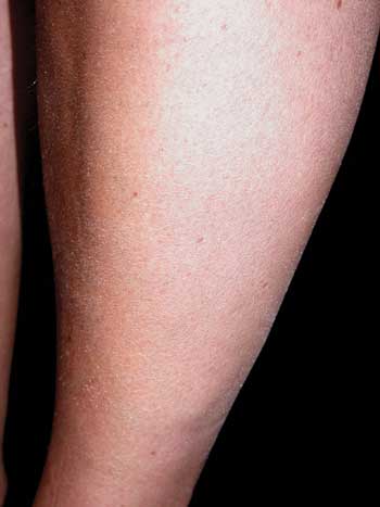 Figure 1: Fine-lamellar scale formation at the lower leg at light-grade Ichthyosis vulgaris |
False Diagnosis 'Dry Skin'
'Dry' in general signifies a lack of moisture respectively water. On the reverse, 'dry' skin would consequently only need moisture in order to normalize again. This conclusion, however, is definitely wrong, as 'dry' skin not only implies a lack of moisture but also of lipids. Insofar we dermatologists should make an effort to designate this skin condition by a non-ambiguous nomenclature and denominate it rather as dry and poor of lipids or as skin poor in hydrolipids. By doing so we are giving a clear signal for an appropriate selection of dermatics and cosmetics in our special field, in the media, in cosmetology as well as towards consumers and patients.
Caused by the imprecision of the term 'dry' skin, quite often over or false care dermatoses are generated. Patients with perioral dermatitis treat their 'dry' skin, which subjectively often produces a feel of tension, with creams rich in lipids, and enhance thus the problem. 'Dry' skin is frequently not treated by persons affected due to the prejudice not to suppress a possible self-production of dermal lipids. Such misinterpretations are able to trigger acute eczema attacks at persons suffering from neurodermatitis.
Skin Poor in Hydrolipids
If avoiding a further use of the term 'dry' skin we achieve a new understanding as well as a higher attention for the 'message' of skin poor in hydrolipids. It is characterized by a reduced activity of the sebaceous gland, a diminished water-binding capacity and/or a disturbance of the stratum corneum structure [1]. Beyond the own sensorium it can be objectified by means of biophysical measuring methods. Thus by using the Corneometer® the hydratation of the horny layer and by means of the Sebumeter® the lipid level at the skin surface can be measured. Parameter such as scaliness, roughness, loss of turgor and wrinkling can be analyzed by Profilometry [1,11]. Far more sophisticated and therefore in the first place reserved for scientific questions is the determination of the trans-epidermal water loss (TEWL) by the Tewameter® [12].
The exact ethiopathogenesis of skin poor in lipids is unsolved. As concerns persons with atopical diathesis, a defect of the function of the stratum corneum barrier is the case, which is able to entail an increased trans-epidermal water loss. Mainly the corneum strateum lipids are lacking [2-4]. A reduced water-binding capacity may be caused by a sebostasis (reduced sebaceous gland activity) [5]. Besides the atopical dermal diathesis as well as with minimal to maximal variants of ichthyosis forms, skin poor in hydrolipids is in general also the case at persons suffering from kidney insufficiency and with diabetics [9,10]. Frequent washing procedures without subsequent skin care may enhance the skin condition poor in hydrolipids in direction of an exsiccation eczema. This particularly applies to risk groups.
The skin of infants is still relatively poor in lipids as the hormonally controlled sebaceous gland activity is not yet fully developed (figure 2). In contrast, the skin is becoming poorer in hydrolipids with ageing, due to the fact that both the sebum production and the quality of epidermis lipids are reduced. At first the areas poor in sebaceous glands at the lower leg are particularly affected by these endogenously age-conditioned changes. Furthermore, based on the deteriorating synthesis of the natural moisturizing factors, the hydratation of the horny layer is reduced [6]. The lipid synthesis is diminished in chronically light-damaged skin, whereas above all the cholesterol synthesis decreases [14].
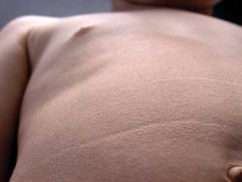 Figure 2: Fine scales at dull-appearing skin poor in hydrolipids at the body stem of a child with clinical symptoms of Ichthyosis. |
Occupational Noxae
The hydrolipid layer of skin is stressed by occupational activities, which are connected with intense humid work. Particularly affected are for example persons employed in the hairdresser's trade, in construction, the metalworking industry as well as in the health sector. Caused by continuous contact of skin with water or aqueous solutions of potential irritants besides the sebaceous gland lipids enriched at the skin surface also the lipids of the strateum corneum can be dissolved from the skin. An impairment of the skin barrier comes about with an increase of the trans-epidermal water loss.
Clinically there is an image at which all characteristics of eczema may coincide (figure 3): skin poor in hydrolipids, maceration, reddening, weeping, roughness, small blisters, chaps and crust formation. Subjectively a more or less intense itching, burning, a reduction of the capability for tactile sensation appears and at times even pain. Moreover, due to the disturbed barrier and reduced resistance function of the epidermis, there is an increased incidence for infections and contact sensitizations.
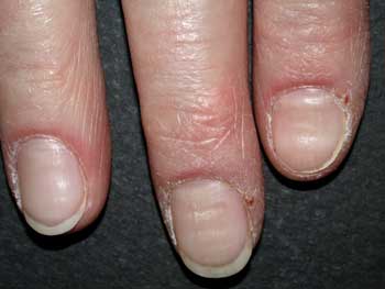 Figure 3: Reddening, scale formation and sebostasis at a patient's fingers with psoriatic and atopical dermal diathesis as well as strong strain by humid work. |
Permeability Barrier
and Epidermis Lipids
The lipids synthesized by corneocytes are accumulated during the epidermal differentiation in the lamellar bodies of the stratum granulosum and conveyed in the intercellular area and hydrolized [1] at the marginal layer (stratum compactum). The lipid fractions thus formed, consisting of cholesterol (20-25 %), ceramides (40 %) and free fatty acids are accumulated lamellar-like at the stratum corneum and significant for the barrier function of the skin [5,7] alike the brick-like structure of the stratum corneum.
It could be shown experimentally that repair processes in the keratinocytes are triggered after artificial disturbances of the permeability barrier. Thus by stimulating of the lipid synthesis the missing dissolved lipids [1] are replaced. By impairment of the stratum corneum, an increase of the TEWL can be initiated. The cause for this phenomenon is not a reduction of the water-binding capacity of the horny layer but a disturbance of the barrier structure based on qualitative or quantitative changes in the composition of the epidermal lipids.
Urea as Natural
Moisturizing Factor
The natural moisturizing factors of skin (NMF) are selectively localized in the stratum corneum and contribute to the suppleness of the horny layer. They substantially consist of various sugars, amino acids, urea, lactic acid and ?-pyrrolidone carbonic acid derivatives [1].
The urea located in the epidermis originates from sweat and the keratisation process. Its water-binding capacity takes place both intra- as well as extra-cellular. In the same way as the other NMF, urea binds water in the horny layer and contributes thus to avoid an excessive trans-epidermal water loss at the transit of skin to the environment.
After an external application urea permeates the horny layer to the corium without a degradation taking place. In dependence of the vehicle and applied concentration, urea is able to trigger besides its water-binding effect also penetration-enhancing, kerato-plastic as well as antipruritic effects. The substance is atoxic and well tolerable at normal skin. When applying it at eczematized skin, however, irritations are caused in general [8].
Therapy and Care of
Skin Poor in Hydrolipids
The therapy and care of skin poor in hydrolipids represents a particular challenge in view of the patho-physiological context described. Preparations recommended by dermatologists should be characterized by hypo-allergenicity, long-lasting effectiveness and an excellent cosmeto-esthetical quality at acceptable price. In order to appear subjectively pleasant for the consumer, the preparations should be well absorbed by the skin and restore the dermal suppleness. Moreover, they should neither leave a fatty gloss nor an unpleasant skin feel. Also odor and the perceptible nature should not be underestimated for the compliance. What possibly functions during a treatment and is accepted in the clinic by the patient may be ceased at home.
In addition, the special properties of the respective base are to be considered for the selection of a preparation. Thus, water-containing preparations with lipid parts of below 20 percent are not suited for the care of skin poor in lipids. Such hydrophilic preparation for topical application have on the one hand the pleasant property to be swiftly absorbed by the skin, are however in general not in the position to normalize an increased TEWL level. In the worst case they are even able to cause an increase of the anyway increased TEWL [1] by the "wick effect". In reverse also mere lipophile bases as lipid ointments or vaseline are only suited to a minor extent for the normalization of the skin poor in hydrolipids as they reduce the TEWL on the one hand but do not contain any moisturizing substances for the improvement of the water-binding capacity.
In order to improve the water-binding capacity of the horny layer, the application of urea-containing preparations for topical application are recommendable. In this context, urea-containing prescriptions in larger quantities (example: urea pura 10 percent in basis cream DAC) are often more cost-effective whereas the advantages of ready-to-use products (for example Elacutan® ointment) are based on the constant production quality and the increased stability. Based on the examinations of the water marked with tritium, urea leads in o/w emulsions to an accumulation of high water quantities in the horny layer already shortly after the application, which however quickly decrease [13]. If however urea is applied in the form of w/o emulsion, the water storage is effected more slowly, lasts however for a longer period of time. From these insights a strategy can be derived for the therapy of the skin poor in hydrolipids to start the treatment with an o/w emulsion and to continue with a w/o emulsion [1].
Besides the hydrophilic phase of the emulsion system, the lipophile phase should equally be arranged in a way that it comes up to the specific requirements of the skin poor in hydrolipids. A preparation for topical application consisting of cholesterol, ceramides and free fatty acids is obviously able to improve the structure of age-caused skin poor in hydrolipids by integration of these fractions into the horny layer barrier [14]. Further, for the product selection localization-typical aspects should be considered. Thus, for example an ideal formulation for the facial skin does not necessarily have to be suited for an application at the palms. The palms due to the lacking of sebaceous glands and a tenfold less concentration of epidermal lipids are considerably lower in lipids than the facial skin. As a result, hydrophilic substances overcome much more easily the skin barrier at the palms than in the face, whereas in reverse lipophile substances in general penetrate the stratum corneum of the facial skin more easily than that of the palms.
Concluding, we dermatologists as specialists for the health and beauty of skin should understand it as a challenge to turn a dull skin poor in hydrolipids and therefore possibly appearing older into an esthetically satisfactory silk-glossy and fully functioning skin surface and thus effectively prevent certain skin diseases.
References
[1] Schürer N, Kresken J: Die trockene Haut – Pathogenese, klinische Manifestationsformen, Probleme der Differentialdiagnose, Galenik, medikamentöse Möglichkeiten, Kosmetika. Wissenschaftliche Verlagsgesellschaft, Stuttgart (2000)
[2] Imokawa G, Abe A, Jin K et al.: Decreased level of ceramides in stratum corneum of atopic dermatitis – An etiologic factor in atopic dry skin? J. Invest. Dermatol. 96 (1991) 523-526
[3] Di Nardo A, Wertz P, Gianetti H et al.: Ceramide and cholesterol composition of the skin of patients with atopic dermatitis. Acta Derm. Venereol. 78 (1998) 27-30
[4] Melnik B, Hollmann J, Plewig G: Decreased stratum corneum ceramides in atopic dermatitis individuals – a pathobiochemical factor in xerosis? Br. J. Dermatol. 119 (1988) 547-549
[5] Elias PM, Grayson S, Lampe MA et al.: Stratum corneum, in Marks R, Plewig G (Hrsg.): The intercorneocyte space, S. 53-67. Springer-Verlag, Berlin, Heidelberg, New York (1983)
[6] Rawlings AV, Scott IR, Harding CG et al.: Stratum corneum moisturizing at the molecular level. J. Invest. Dermatol. 99 (1994) 731-740
[7] Schürer N, Elias PM: The biochemistry and function of stratum corneum lipids. Adv. Lipid Res. 24 (1991) 27-56
[8] Hornstein OP (Hrsg.): Externe Therapie von Hautkrankheiten. Thieme-Verlag, Stuttgart, New York (1985)
[9] Morton CA, Lafferty M, Hau C et al.: Pruritus and skin hydration during dialysis. Nephrol. Dial. Transplant. 11 (1996) 2031-2036
[10] Altmeyer P, Kachel HG, Jünger M et al.: Hautveränderungen bei Langzeitdialysepatienten. Hautarzt 33 (1982) 303-309
[11] Rohr M, Schrader K: Fast optical in vivo topometry of human skin (FOITS) – Vergleichende Untersuchungen zur Laserprofilometrie. SÖFW 2 (1998) 3-8
[12] Barel AO, Clarys P: Study of stratum corneum barrier function by transepidermal water loss measurements – Comparison between two commercial instruments: Evaporimeter® and Tewameter®. Skin Pharmacol. 8 (1995) 186-195
[13] Wohlrab W: Harnstoff – ein bewährter Wirkstoff in der Dermatologie und Kosmetik. Pharm. Ztg. 137 (1992) 2483-2489
[14] Zettersten EM, Ghadially R, Feingold KR et al.: Optimal rations of topical stratum corneum lipids improve barrier recovery in chronologically aged skin. J. Am. Acad. Dermatol. 37 (1997) 403-408
| Authors'
Address: Dr. med. Erik Senger Hautarztpraxis Breidertring 104 D-63322 Rödermark E-Mail:erik.senger@t-online.de |
top